NeurologyDra. Beatriz Chavarría Cano
Dr. Javier Emilio Codas Campuzano
Dr. Gabriel Salazar Tortolero
| Neurosurgery
Dr. Cristóbal Perla y Perla Fuentes Dr. Juan Camilo Hernández Acevedo
Dra. Mireia Illueca Moreno
Dr. Agustín Bescós Cabestre
Dr. Fran Martínez Ricarte
Dr. Esteban Cordero Asanza
|
Neurophysiology
Dra. Estela Lladó Carbó
Dra. Maria Eugenia Vera Pavón
Dra. Raidili Mateo Montero
| InterventionalNeuroradiology
|
Sleep Study unitDr. Rodrigo Rocamora Zúñiga
Dra. Raidili Mateo Montero
|
Epilepsy
Dr. Rodrigo Rocamora Zúñiga
Dra. Beatriz Chavarria
Dra. Carmen Pérez Enríquez
Dr. Jaume Capellades Font
|
Comprehensive Oncology Unit
HM CIOCC
At the new Clara Campal Comprehensive Oncology Center (HM CIOCC) HM CIOCC-Barcelona, our commitment is to offer quick and personalized attention that guarantees each patient the optimal diagnosis and treatment for their disease.
We assess each patient in specialized multidisciplinary Medical Committees for each type of cancer.
Each committee has surgeons and oncologists with proven experience, as well as other specialties involved in the approach to cancer. An accessible team that continuously incorporates therapeutic advances. At HM CIOCC Barcelona we also develop an innovative networking that ensures quality and innovation in cancer treatment.
Medical Director: Dr. Joan Albanell Mestres
Radiology Service
In addition to conventional and remote-controlled radiology devices, the Diagnostic Imaging Service has:
• 160 multislice CT scan (Toshiba Aquilion) with 80 detectors. It can perform coronary artery examinations and the most complex studies in a few minutes.
• Closed high field MRI (RNM MAGNETOM ESPREE Siemens 1.5 T) - High magnetic field equipment with a compact magnet that is wider and shorter than conventional resonances. Indicated for claustrophobic and obese patients, it provides high quality images.
• Breast Pathology Unit. In this unit, we offer a digital mammogram with tomosynthesis (SELENIA DIMENSIONS). We have also other systems for advanced imaging such as sterotaxy, and the latest generation breast ultrasound (ESAOTE MyLabEight XP), an ultrasound system that defines a new standard in image quality for a better diagnosis.
• EOS – a low radiation 3D vertical radiology: Vertical radiological diagnostic system that allows obtaining 2D images and 3D reconstructions, having the patient standing up or even sitting in a transparent chair to X-rays.
• Prostate Fusion Biopsy: Currently, it is the most effective technique for the diagnosis of prostate cancer thanks to the combination of images obtained by conventional transrectal ultrasound and those of magnetic resonance, a prostate mapping is generated. This information is very precious for the specialist when the biopsy is made. In this way, samples are collected only from areas detected as tumorous or suspicious.
• PHILIPS ECOGRAPH (AFFINITY 70 DS): Conventional ultrasound (muscle, transference, soft tissue…)
• PORTABLE ECOCARDIO (PHILIPS COMPACT EXTREM CX-50 CARD)
• GYNECOLOGICAL ECOGRAPHY (GENERAL ELECTRIC-VOLUSON 730 Pro): Abdominal/vaginal gynecological ultrasound and 3D ultrasound.
• ORTHOPANTOMOGRAPH (ORTHORALIX 9200 GENDEZX): For dental X-rays and skull X-rays.
• FIBROSCAN (FIBROSCAN PROBE M +) - Study of the elasticity of the liver (fibrosis fracture) replacing liver biopsy.
Radiology - Diagnostic Imaging
The Diagnostic Imaging Service of HM Delfos Hospital is one of the pioneers in implementing and disseminating radiological techniques in Spain, having the latest digital technology in Diagnostic Imaging.
Thanks to Radiology we can generate images of the inside of the body for a better diagnosis and treatment of diseases.
This Service performs ultrasounds, digital mammography, EOS - Vertical 3D low radiation radiology system, contrast radiology, dental radiology, magnetic nuclear resonance and CT, among other tests.
This Service covers Interventional Radiology with techniques and procedures such as the placement of prostheses, catheters and drains by percutaneous route, puncture biopsy, puncture and drainage of directed collections, hepatic tumoral chemoembolization and percutaneous vertebroplasty, among others.
In addition, the Diagnostic Imaging Service of HM Delfos is a pioneer in carrying out 3D studies in Spain, and currently carries out 4D ultrasound scans as a technique that allows better vision of the fetus or vertical 3D X-rays (EOS) with ultra-low radiation dose.
Highest level of technology
In addition to conventional and remote-controlled radiology equipment, the Diagnostic Imaging Service has
• 160 multislice CT scan (Toshiba Aquilion) with 80 detectors. It can perform coronary artery examinations and the most complex studies in a few minutes.
• Closed high field MRI (RNM MAGNETOM ESPREE Siemens 1.5 T) - High magnetic field equipment with a compact magnet that is wider and shorter than conventional resonances. Indicated for claustrophobic and obese patients, it provides high quality images.
• Breast Pathology Unit. In this unit, we offer a digital mammogram with tomosynthesis (SELENIA DIMENSIONS). We have also other systems for advanced imaging such as sterotaxy, and the latest generation breast ultrasound (ESAOTE MyLabEight XP), an ultrasound system that defines a new standard in image quality for a better diagnosis.
• EOS – a low radiation 3D vertical radiology: Vertical radiological diagnostic system that allows obtaining 2D images and 3D reconstructions, having the patient standing up or even sitting in a transparent chair to X-rays.
• Prostate Fusion Biopsy: Currently, it is the most effective technique for the diagnosis of prostate cancer thanks to the combination of images obtained by conventional transrectal ultrasound and those of magnetic resonance, a prostate mapping is generated. This information is very precious for the specialist when the biopsy is made. In this way, samples are collected only from areas detected as tumorous or suspicious.
• PHILIPS ECOGRAPH (AFFINITY 70 DS): Conventional ultrasound (muscle, transference, soft tissue…)
• PORTABLE ECOCARDIO (PHILIPS COMPACT EXTREM CX-50 CARD)
• GYNECOLOGICAL ECOGRAPHY (GENERAL ELECTRIC-VOLUSON 730 Pro): Abdominal/vaginal gynecological ultrasound and 3D ultrasound.
• ORTHOPANTOMOGRAPH (ORTHORALIX 9200 GENDEZX): For dental X-rays and skull X-rays.
• FIBROSCAN (FIBROSCAN PROBE M +) - Study of the elasticity of the liver (fibrosis fracture) replacing liver biopsy.
Heads of Radiology - Diagnostic Imaging
Dr. Josep Maria Argilés Vives
Dr. Josep Vives Roura
Radiology - Diagnostic Imaging
Monday to Friday: 8:00-20:00
• 24h / 365 days TAC Emergency Service
HM Delfos hospital incorporates the most technologically advanced CT with ultra-fast 3D reconstruction capabilities
Barcelona, November 5th, 2018. The Hospital HM Delfos in Barcelona has incorporated a new 160-crown CT scan that obtains high quality images in a very short time and will allow more than 24,000 examinations per year.
This new generation CT, with a high efficiency and low persistence detector tray, is one of the most advanced technology CTs in Barcelona's hospitals, as it allows ultra-fast reconstruction with 3D functions.
In this way, HM Hospitales is once again at the forefront of the adoption of healthcare technology aimed at offering its patients and medical teams the most advanced tools in order to provide the best healthcare.
More effective and with less radiation
The heads of the Radiology Service of the Hospital HM Delfos, Dr. Josep Vives, as well as Dr. José María Argilés, have explained that "the radiation dose is optimized with advanced iterative algorithms of UltraHelical reconstruction and acquisition. This means that the radiation is reduced to the needs of each patient and each test.
They also pointed out that "the specific software included in this CT minimizes the possibility of repeating the tests, since it makes it possible to detect arrhythmias and determine the cardiac phase. In the same vein, "this technology with algorithms makes it possible to clearly visualize structures that were previously obscured by the presence of metal implants.
Likewise, the use of this latest technological tool helps the medical team at HM Delfos to plan and make the best decisions for patients. "The 3D functions allow for ultra-fast reconstruction, with automatic bone removal and reconstruction," they said.
Continued technology investment in the coming months
HM Hospitales maintains its commitment to technological renovation at HM Delfos with this 160-crown multidetector CT, in addition to the implementation of the only vertical EOS scanner in Spain that allows for 3D images in only 20 seconds of the full column and extremities, reducing radiation to the patient by 85%.
In 2019, HM Delfos will complete this technological investment with the acquisition and implementation of a 3T magnetic resonance imaging system, which will place the HM Delfos Diagnostic Imaging Department on the same level as third-level high-tech public hospitals.
Breast Pathology Diagnostic Unit
Mammography, Ultrasound and Magnetic Resonance
In our Breast Pathology Image Diagnosis Unit, integrated in the Image Diagnosis Service of the Hospital HM Delfos, it includes the study of screening patients in asymptomatic women, the diagnostic study in the presence of symptoms or signs of breast disease and the monitoring of benign pathologies and carcinoma after treatment.
At the forefront of technology
We have the most advanced diagnostic technology with Digital Mammography (with Tomosynthesis) and High Definition Ultrasound, as well as high field closed Magnetic Resonance.
We also have techniques of proven effectiveness in diagnostic breast surgery, with ultrasound guidance and digital stereotactic.
Tumor Committees
We participate in the Tumor Committee of the Hospital HM Delfos, in which all the specialists involved in the diagnosis and treatment of breast cancer participate, which guarantees coordinated, planned and comprehensive care of breast pathology in all its aspects: clinical, diagnostic and therapeutic.
Our portfolio of explorations
• Digital Mammography
• Tomosynthesis
• Breast Ultrasound
• Axillary ultrasound
• Closed MRI 3T
• Cytological puncture
• Thick needle puncture
• Core needle biopsy with ultrasound and stereotaxy
• Tumor Marker test
• Locating nodules with a computed axial tomography-guided harpoon
Head of the Diagnostic Unit of Mammary Pathology
Dr. Montserrat Clotet Feliu
Hospital HM Delfos incorporates a state-of-the-art mammography machine
Barcelona, November 14th, 2018. The Hospital HM Delfos in Barcelona has incorporated a state-of-the-art mammography machine that will substantially improve the comfort and speed of the test and the results obtained for the patients.
This mammograph with 3D Tomosynthesis "improves the definition and therefore generates more confidence in the reading of the tests by radiologists" assures Dr. Montserrat Clotet Feliu, specialist in Radiodiagnosis of Mammary Pathology of the Hospital HM Delfos. In addition, she adds that "excellent optimization of mammography images is achieved, acquiring them more quickly. In this way, the speed in obtaining high quality images from the new mammograph will allow between 8,000 and 10,000 explorations to be carried out per year.
The patients will have to spend less time for the test and, therefore, it will be more comfortable for them. In addition, the new mammograph contributes to the earlier detection of invasive malignant lesions and reduces the number of recitals by 40%.
One case every 20 minutes
Every 20 minutes an oncological process of this type is diagnosed in Spain, which results in 26,000 new patients per year. Therefore, it is essential that women with breast cancer have an active role in making decisions about their disease and, of course, in everything related to clinical research.
HM Hospitales insists on not letting down its guard with prevention - the help of the new mammograph is vital for doctors and patients - and to react quickly to new findings in the breasts: lumps, skin changes, retraction or bleeding from the nipple.
With the arrival of the new mammography machine, HM Hospitals continues with its innovative investment plan aimed at placing the group's new center in Barcelona among the best in terms of healthcare technology adoption.
This is the third major innovation in less than a month at HM Delfos after incorporating a 160-crown CT with ultra-fast 3D reconstruction functions and the EOS, a vertical radiology system that makes it possible to obtain an image in just 20 seconds, does not generate claustrophobia and its radiation is up to 85% less than traditional X-ray systems.
HM Delfos Hospital continue its technological and physical transformation to become a reference in the Catalan private healthcare system.
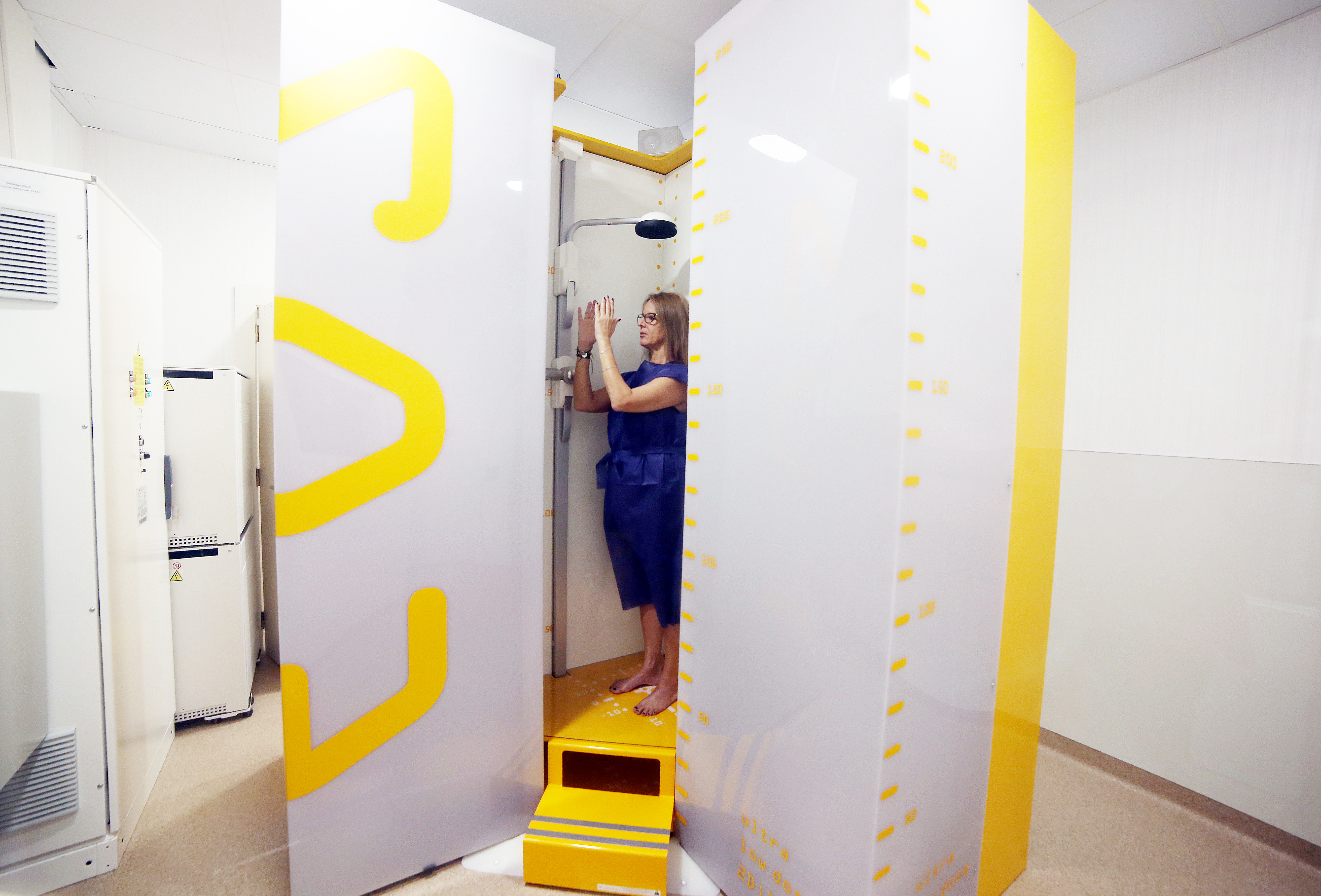
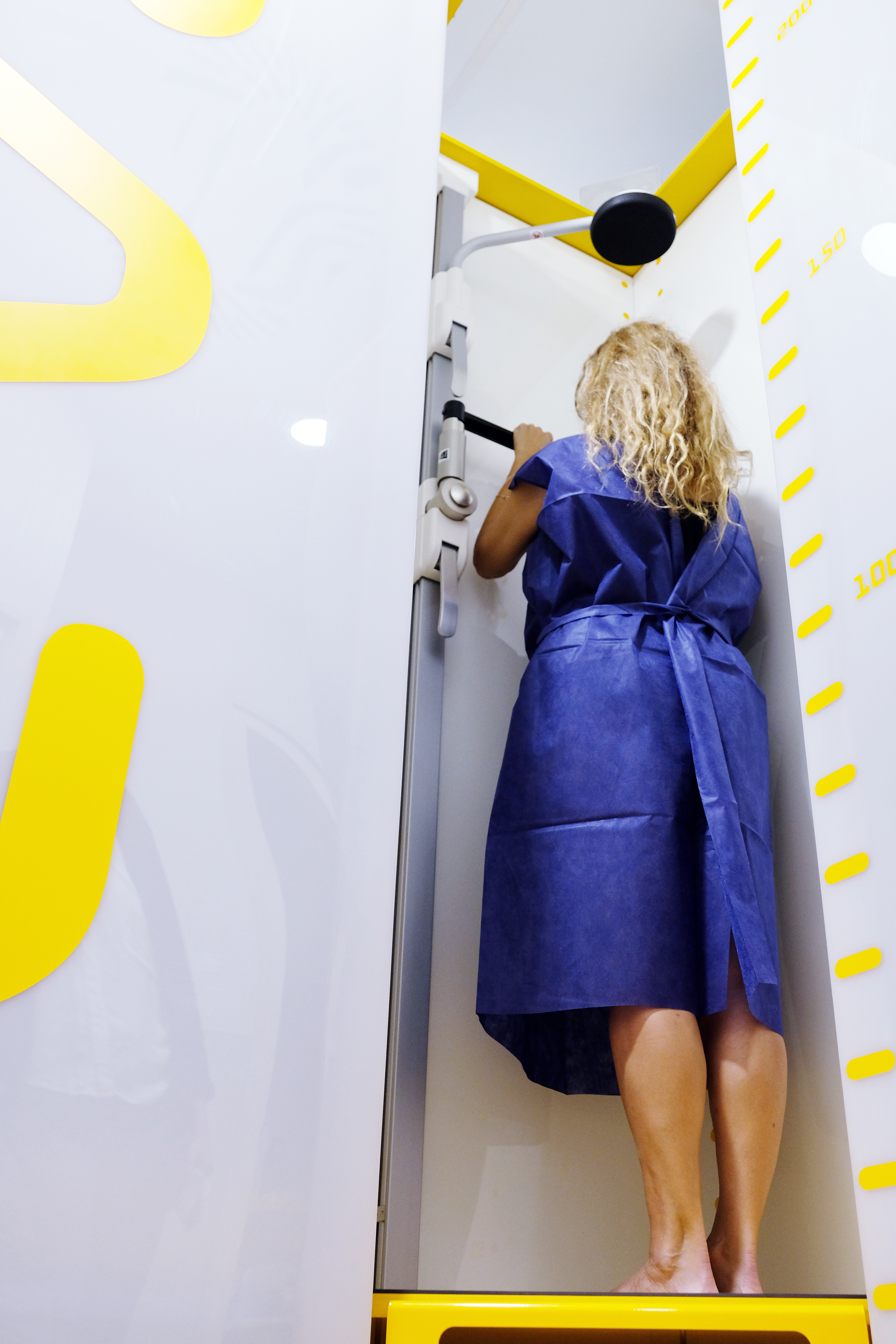
EOS - Low Radiation Vertical 3D Radiology System
HM Delfos, the first hospital in Spain to offer the EOS® 3D diagnostic system with low radiation.
EOS
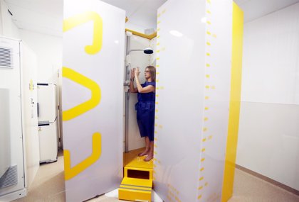
What is EOS?
It is an image diagnosis system that allows to obtain images of the whole body in traditional projections (2D) and in 3D reconstructions, where the patient is found in a loading position, that is, sitting in a transparent chair, the X-rays or standing up.
By means of the two full-body images, it is possible to create 3D models of the super skeleton. The specialist will use the measurements and data from the 3D model to make a diagnosis and prepare a personalized treatment for you as part of the shared decision making process.
This technology is dedicated to adult and child patients with musculoskeletal pathologies.
Benefits for the patient
Less radiation
One of the main features offered by EOS® is that it allows for better diagnoses with less radiation. The current alternative to EOS® is a CT scan, but in this test the patient is stretched, without carrying his own weight, and the radiation is 85% higher. These features make it possible to make a better diagnosis, monitor a patient's progress more reliably, or be able to accurately plan surgery.
This minimal radiation emission can be even more reduced for pediatric patients and/or those who need to undergo multiple controls, since the radiation received is significantly less than that emitted by traditional radiology and CT systems.
The EOS® System has less radiation, 50% less than traditional X-rays and up to 85% less than computed tomography (CT) scans.
"In the case of controls, acquisitions with monodose (85% less radiation) or with even less microdose, which is equivalent to the natural environmental exposure of one week, can be used. This reduction in radiation is especially significant in the pediatric population, being very relevant for complex problems such as scoliosis, in which a series of body images must be obtained repeatedly over time.
This reduced radiation is due to a system of particle emission and X-ray detection, a system which earned its inventor, Georges Charpak, the Nobel Prize for Physics in 1992.
It does not generate claustrophobia
In addition, the wide space of the cabin does not generate claustrophobia.
High image quality
The image is generated by a simultaneous double vertical scan, front and profile, which lasts about 20 seconds for the whole body. By capturing the exams in a vertical, weight-bearing position, specialists can
• Better assess the patient's overall posture
• Understanding the relationship between the spine, pelvis and lower extremities
• Understand the compensatory mechanisms of the lower extremities
During post-processing, 3D images can be generated, but it is possible to go further, since it allows the data to be entered into a surgical planning program to transfer the results obtained with the EOS® to the operating rooms. This planning is possible because the measurements made with the EOS® System are distortion-free and very precise.
This additional data is essential because specialists improve diagnosis and treatment decisions and plan surgical interventions with better performance, allowing for improved patient outcomes.
For which pathologies is EOS® indicated?
EOS® is especially indicated for detecting problems in skeletal and joint pathologies, as well as for degenerative diseases of the spine, knee and hip. The main reason for the orientation towards these pathologies lies in the possibility of obtaining high quality images in load. Full-body 3D imaging is also important for the evaluation of many other specialties besides spine surgeons. These images complement biomechanical studies, and are very important in specialties such as Podiatry, Orthopedics, Traumatology, Rheumatology, Rehabilitation and Physiotherapy, Osteopathy and Neurosurgery.
The EOS® vertical scanner improves the quality of life of children who need periodic image controls
Improvement for patients at HM Children's Hospital
The main beneficiaries of this new technology will be the pediatric patients of the Hospital HM Nens de Barcelona, which has recently joined the Group's healthcare network in Barcelona, where it also forms part of the Hospital HM San Jordi in the San Andrés district. Dr. Gerardo Conesa, director of the Comprehensive Center for Neurosciences - HM CINAC Barcelona, located at HM Delfos, explains that "from now on, when faced with a pediatric patient suffering from a skeletal-type disease that requires us to perform frequent radiological checkups, as is the case with scoliosis, we will no longer have to be so demanding when assessing whether or not a test is strictly necessary for fear of subjecting the patient to excessive radiation, because with EOS® the level is very low.”
Scoliosis, a major reason for consultation in Pediatrics
The idiopathic scoliosis is a deformity of the spine superior to 10⁰ that teenagers especially suffer. As prevention, it is recommendable that the specialist explores the minors regularly as of the 10-11 years. Nowadays, the prevalence of this disease is 2% and is one of the main reasons for consultation in the field of Pediatrics. The head of the Diagnostic Imaging Service at HM Nens Hospital, Dr. Ramon Delgado, explains that "there are many patients that we visit in our center because of this and other back pathologies, so being able to have EOS® technology now means a great advance for us, since we can offer a much more precise diagnosis in less time and, in the case of having to carry out a treatment, we can make a more rigorous follow-up, since the children receive much less radiation than with telemetry or CT".